Bio class travels to UCSD to perform endoscopies on pigs
Pig stomachs shine a light on human stomach disorders
March 25, 2015
By Mati Davis, Arts & Culture Editor
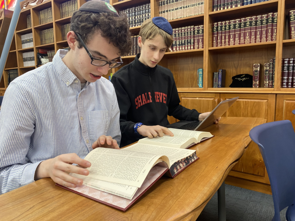
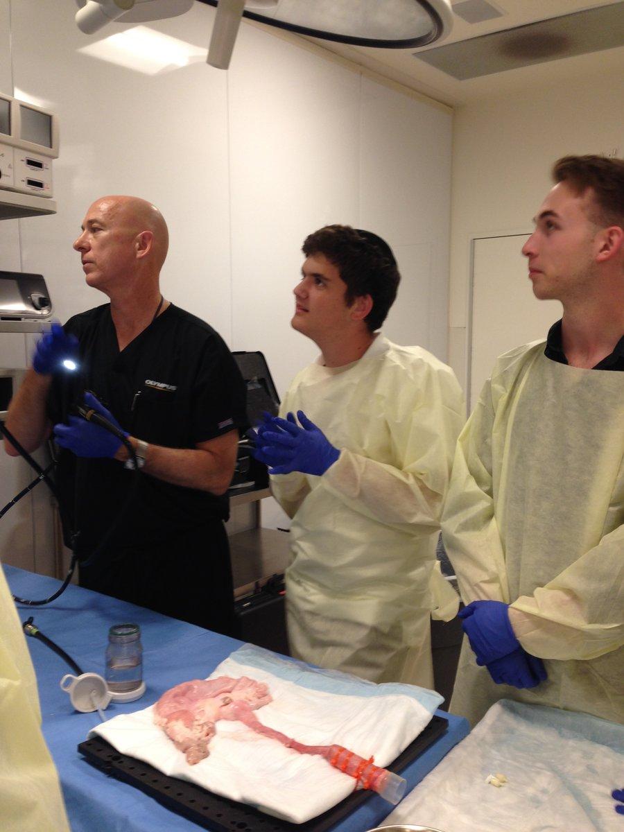
Have you ever wondered what it’s like to look inside a pig’s stomach? Seven Shalhevet juniors and two seniors joined students from YULA, B’nos Devorah and Yavneh and traveled by bus to UC San Diego Feb. 24 to learn about Inflammatory bowel disease and perform endoscopies on pig stomachs.
The stomachs had been removed from the animals in advance.
“At first it was a little startling, but it was just the stomach, so it didn’t look like a pig,” said junior Rose Lipner. “By the end of the endoscopies the pig stomach was sort of a separate entity, and didn’t really have any association with an animal.”
An endoscopy is a procedure in which a tube with a camera at the tip is insert down the throat into the esophagus, and from there into the mucus-filled stomach and small intestine to look for signs of Crohn’s disease, ulcerative colitis and other conditions. Students took turns maneuvering the machine’s camera, with the image displayed on a monitor above the table.
The bus ride south had been around two hours. At around noon, students arrived at UCSD’s Imflammatory Bowel Disease
Center, where after much waiting the roughly 60 students high schoolers headed downstairs to a lab and were given yellow surgical scrubs and surgical gloves. They left all belongings in a locker room.
Students were then led into a room with six surgery tables, each with a deflated pig stomach and duodenum. The students were then divided into six groups of approximately seven people per group and professional scientists led them through the endoscopy process.
Junior Daniella Banafshea especially enjoyed this part.
“I was able to learn about the illness and how to do a endoscopy,” said Daniella. “The most fascinating part was doing the endoscopy myself.”
University staff even let the students play a game, inserting small objects into the stomach and having the students take a special tool, used with the endoscope, to remove them.
The students were very nervous as they took the controller in their hand, trying to weave the endoscope through the veiny muscles of the pig stomach. Once past the esophagus, the rest of the group cheered and encouraged the endoscoper.
“It was really cool to see what an actual stomach looked like even if it was just a pig,” said junior Kayla Ablin. “It was also interesting that we got to use the same tools doctors use when they do endoscopes on humans.”
Before the endoscopy, the students attended a lecture on a the describe the effects of IBD. Later a professor from UCSD explained its causes.
“We got a lot more than we thought we were getting,” said junior Daniel Shoham. “I originally thought we would just be learning how to perform an endoscopy, but I found it interesting to in addition learn more about IBD and Crohn’s disease”.
The students from the different schools stayed together throughout the program, eating lunch, answering questions in the lecture hall, and riding to and from the campus’s grassy area. All the Shalhevet students sat in a circle sharing stories and telling jokes.
Biology teacher Dr. Melissa Noel said the trip helped helped fill the gap caused by Shalhevet’s building being under construction.
“We haven’t had the opportunity to do any labs this year, so this was the student’s opportunity to do hands-on work,” said Dr. Noel.
At approximately 4 p.m., everyone boarded the bus and rode for three hours back to Los Angeles.
“It was fun to do some hands-on work and really learn about endoscopy in a fun, interactive way,” said junior Yonah Feld.